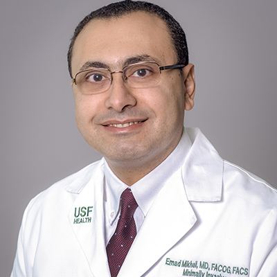
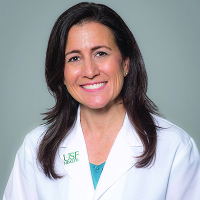
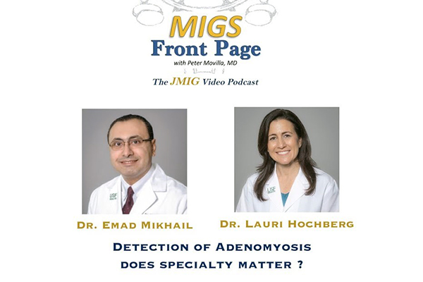
USF Health Morsani College of Medicine physicians Emad Mikhail, MD, and Lauri Hochberg, MD, were recently featured in a Journal of Minimally Invasive Gynecology video podcast about how pelvic ultrasound detection of adenomyosis, a benign condition causing thickening of the uterine wall, may be affected by the specialty of the sonographic provider.
Drs. Mikhail and Hochberg, both assistant professors, were among the MCOM Department of Obstetrics and Gynecology faculty members who conducted the study appearing online in Journal of Minimally Invasive Gynecology in April 2019 and highlighted in the recent podcast. Dr. Mikhail was the research paper’s senior author and Dr. Hochberg, director of USF Health’s Gynecological Imaging Service, was a co-author.
The USF Health researchers found that the preoperative adenomyosis detection rate was significantly higher when the ultrasound was performed by gynecologists with expertise in gynecologic imaging compared to radiologists.
Adenomyosis occurs when tissue lining the uterus grows into the muscular wall of the uterus. While about a third of women experience no symptoms, the condition can lead to an enlarged uterus and painful, heavy periods.

Dr. Emad Mikhail, the research paper’s senior author, specializes in minimally invasive gynecologic surgery.
The single-center retrospective analysis was based on 412 histopathologically confirmed cases of adenomyosis from hysterectomy specimens. After controlling for variables — including race, body mass and prior Cesarean sections, and uterine fibroids– the researchers found that the expert gynecologic sonologists were 7.8 times more likely to identify adenomyosis than the radiologists. The presence of fibroids and uterine enlargement significantly decreased detection by both specialty groups.
Dr. Mikhail noted that although the expert gynecologic sonologists had a detection rate more than 7 times higher than radiologists, they still only identified adenomyosis 56 percent of the time (versus 26 percent for radiologists).
“We need to improve our detection rate of adenomyosis among symptomatic patients he said. “Overall, I think this is a thought-provoking finding that will help provide ideas for future studies.”

At USF Health Dr. Lauri Hochberg directs one of few practices across Florida certified in 3D gynecologic ultrasound by the American Institute of Ultrasound in Medicine.
The need is growing for practitioners with expertise in gynecologic imaging to improve diagnostic accuracy for clinically complex conditions like adenomyosis, said Dr. Hochberg, who is certified in 3D gynecologic ultrasound by the American Institute of Ultrasound in Medicine (AIUM). She pointed to a recent AIUM consensus report calling for standardized curriculum and competency assessment tools to be used by residency programs for obstetric and gynecologic ultrasound training.
“I think that real-time gynecologic imaging while the patient is with you in the exam room, so you can ask how they feel and obtain a medical history, helps us (expert gynecologic sonographers) better understand and detect problems more often… If you’re taking pictures and reading the ultrasound from a different room you may miss a lot.”

